
MRI is no longer optional in urology. As Dr. Edward Matsumoto, urologist at St. Joseph’s Healthcare Hamilton, put it in his conversations with our product team:
“MRI has become a part of everyday life in Urology.”
Dr. Matsumoto orders MRI scans for his patients daily. He uses the highly detailed scan to plan care and surgical approaches, given the clarity only MRI can provide.
And yet, despite MRI’s central role in diagnosis, most interventional prostate procedures still depend on approximations and subpar ultrasound visualization.
That’s the precision care gap.
How the precision gap affects Dr. Matsumoto’s prostate surgery practice
Dr. Matsumoto specializes in minimally invasive prostate surgery, including robotic-assisted prostatectomy. Like many leading urologists, he routinely leads patients through treatment options after a diagnosis, including surgical options. To help patients make the right choice for their health, Dr. Matsumoto needs confidence in biopsy results, and encounters the limitations stemming from today’s biopsy procedures.
When scanned with an MRI, the lesion(s) within a prostate are clear. But when that image is merged using fusion software with ultrasound images to take a biopsy, the urologist is forced to guess.
That’s because fusion software works off the previously captured MRI image to calculate where a lesion should be, rather than showing the urologist exactly where it is. This make-do compromise means the urologist must guess where to place a biopsy needle within the prostate, an experience we’ve compared to trying to hit the bullseye on a dart board while wearing blurry glasses.
This imprecision limits a urologist’s confidence in the biopsy results—and confidence determines whether minimally invasive treatment options are viable. In this paradigm, biopsy results become uncertain, patient eligibility pools for focal therapy get shallower, and, in many cases, patients get pushed toward whole-gland removal.
The issue isn’t a lack of technology. It’s the disconnect between imaging and where its precision is needed most—the intervention
Glenn™: A robotics platform designed to close the precision care loop
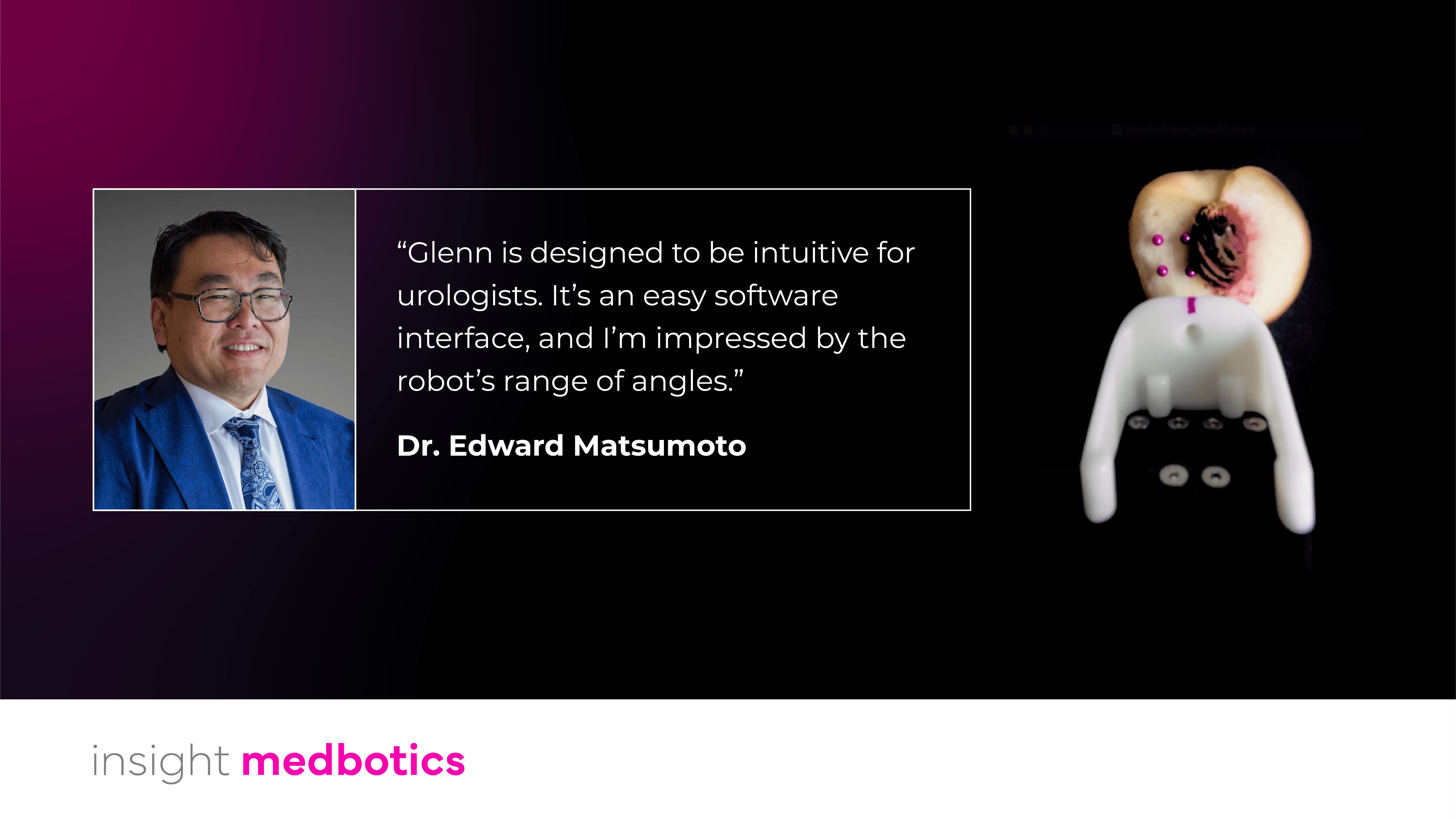
When Dr. Matsumoto saw Glenn™, he immediately understood the shift: “Glenn is designed to be intuitive for urologists. It’s an easy software interface, and I’m impressed by the robot’s range of angles.”
Glenn doesn’t add complexity. It simplifies MRI-guided workflows by closing what we call the Precision Care Loop. By bringing real MRI guidance directly into urologic practice, Glenn creates continuity for urologists and their patients.
Glenn links the three steps that are typically siloed in current practice:
- Diagnosis – MRI-visible lesions are imaged and targeted directly for biopsy, not approximated through fusion.
- Mapping – Every biopsy core is spatially recorded with exact coordinates inside the prostate.
- Pathology and Therapy – Cancer diagnosis results for each biopsy core are mapped to the location where they were obtained from the prostate. With that specific map, those same coordinates can guide focal treatment, ensuring energy is delivered precisely where cancer was confirmed.
Instead of guessing, urologists can see exactly where they are, where they’ve been, and where they’re going across the entire care pathway.
That shift—from approximation to continuity—radically changes what’s possible.
Why continuity matters for the future of prostate cancer care
Focal therapy is the logical next step in minimally invasive prostate cancer treatment. But adoption has been constrained by uncertainty. Dr. Matsumoto can see what happens when that uncertainty disappears:
“On focal, it will be a game changer and may be a challenger to da Vinci robotic prostatectomies.”
That’s not to dismiss surgical robotics, but more to recognize that as upstream precision improves, treatment decisions evolve with greater certainty. Whole-gland prostate removal runs the risk of life-altering side effects. Closing the precision care loop builds the certainty required to clear more patients for focal therapy, which will in turn decrease the number of patients with no recourse beyond whole prostate removal.
MRI has already transformed how prostate cancer is diagnosed. The next shift is making that same imaging power fully accessible to urologists during intervention, without adding friction, complexity or workflow disruption.
We’re designing Glenn to make it easier to shorten procedure time, automate trajectory alignment, and integrate with existing MRI systems. We want to make interventional MRI more practical for hospitals that balance scanner use between diagnostics and interventions.
And, as Dr. Matsumoto notes, when it comes to adoption: “easy is good.”
—Glenn™ is under development for prostate applications and not yet FDA cleared for that indication in the U.S.